
by Tammy Stearns, MS, RDMS, RVT, RT(R), FSDMS, FAIUM, LAS
Screen orientation plays a significant role in our ability to image well. When the imager understands screen orientation, this allows for recognition of the images on the screen and significant landmarks. If an imager is scanning and loses sight of the screen orientation, it is best to start over. For example, return to the place that is known such as midline uterus or another landmark that is familiar. If helpful, the imager can pick the transducer up and reset it at the level of the pubic symphysis. Then, start completely over instead of struggling and trying to find something that is recognizable and getting continually frustrated. While it is noted that screen orientation is one of the most basic concepts of scanning, it is also to be noted that once this concept is mastered, the imager’s ability to create images improves significantly as they are able to understand and manipulate the sound waves to image with greater clarity and confidence.
It helps to keep in mind that the face of the transducer corresponds to the first structure that the sound wave comes into contact with. Transabdominally, that is the gel and then it is the client’s skin. Most often transabdominally, this point of the transducer, which will appear at the top of the screen of the ultrasound image, will coordinate with the anterior abdominal wall. The image continues to be built utilizing the go-return-time of the sound waves by placing each pixel of gray depending on the amount of time it takes for the sound wave to leave the transducer and interface with a structure and return to the transducer. The longer the time, the further down on the screen the pixel of gray is placed. This is a point where confusion tends to set in. Since the pixel is placed lower on the screen, oftentimes, it is misinterpreted as being inferior when it is actually posterior in the body.
The same concept can be utilized when explaining transvaginal screen orientation. The face of the transducer is placed in the vagina and interfaces first with the cervix placing the top of the screen as inferior and the bottom of the screen as superior. Again, it is helpful to remember the first structure the sound wave comes into contact with will correspond to the top of the ultrasound image. The last structure that it comes into contact with will correspond to the bottom of the image. The other pixels of gray lie in-between depending on how long it takes for them to travel from the transducer to the structure and back.
Looking deeper into the orientation, the notch corresponds to the right side of the ultrasound screen. This is why when the notch gets inadvertently turned into another direction it can seem as if the screen and transducer are not in alignment. When scanning transabdominally in a longitudinal plane, pointing the transducer notch toward the patient's head ensures that the left side of the screen displays superior structures (toward the head) and the right side displays inferior structures (toward the feet). When performing a transvaginal ultrasound in the longitudinal plane, orient the transducer notch toward the ceiling. This aligns the right side of the screen (the notch side) with the patient's anterior anatomy, and the left side of the screen with the patient's posterior anatomy.
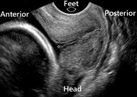
Transvaginal ultrasound screen orientation, longitudinal.
These orientations hold true when in the longitudinal plane of scanning with the notch turned either towards the client’s head (transabdominally) or towards the ceiling (transvaginal). When we move to the transverse plane, with the notch towards the client’s right (both transabdominally and transvaginal), the orientation of the right and left sides of the screen change because the part of the transducer that corresponds with the notch has changed direction of orientation. The part that the face of the transducer is in contact with has not changed, in transabdominal and the top of the screen still corresponds anterior with the bottom of the screen posterior. In transvaginal, the top of the screen still corresponds inferior and the bottom of the screen is superior. The only time when this part of the orientation will change is when we change where the face of the transducer first comes into contact.
When the transducer is turned transverse, the machine is still calibrated to associate the notch side of the transducer with the right side of the screen. Transabdominally, the right side of the screen (client right) will correspond to the right side of the client. The left side of the screen (client left) will correspond to the left side of the client. This is true transvaginally also.
As we mature in our scanning process, we will begin to memorize concepts and we will start to understand how the art of imaging is more than just memorization. The art of imaging is learning to be able to put physics concepts, intertwined with structural anatomy, together to create an image that not only gives our client important information, but also creates an image that makes a lasting imprint on their lives. As we continue to practice and hone our skills as an imager, utilizing it more than just a medical exam and remembering that we are indeed witnessing Holy Ground as He knits His creation together in its mother’s womb, we will adopt a reverence that will enhance each and every scan that we have the privilege to image.
