
by Tammy Sterns, MS, RDMS, RVT, RT(R), FSDMS, FAIUM, LAS
The Limited Obstetrical Ultrasound Exam performed in Pregnancy Help Medical Clinics (PHMCs) answers specific questions to provide the mother with the information that she needs to make an informed decision regarding her pregnancy. The exam itself is limited in nature due to the narrowed purpose of the exam. Typically, the mother is needing to know the gestational age of the pregnancy along with the approximate due date. By virtue of the integrity of the scan, the imager also seeks to confirm that the pregnancy is, at that moment, alive and is located within the endometrial cavity within the uterus. The imager is also able to visualize the baby so that the mother can actually see the life inside of her and realize the reality of the pregnancy to begin the mother-fetal bonding process. This is what is unique regarding imaging in PHMCs.
Our focus is life-affirming for both the mother and her baby. If the mother chooses to continue the pregnancy, a more detailed, complete ultrasound exam is performed to assess the pregnancy and baby once she begins prenatal care.
The limited scan images the vagina, cervix, uterus, and adnexa in both longitudinal and transverse scanning, the entirety of the structures to provide visualization and thorough evaluation of the pelvic area. It is recommended to perform a systematic scan to ensure that the exam images all the structures. Starting longitudinal at the pubic symphysis, the vagina, cervix, and uterus are imaged midline. The scan continues angling towards the mother’s right side through the uterus and the entirety of the right adnexa. Returning back to midline, the imager angles left through the uterus and the entirety of the left adnexa. Turning the transducer transverse (notch to the mother’s right), the imager, remaining in the sonographic window of the full bladder located at the level of the public symphysis, angles superiorly, noting the level of the vagina, cervix, lower uterine segment, transverse uterus, fundus of the uterus, and angling superior past the uterus. Transverse imaging of the right and left adnexa, scanning from inferior to superior, is also performed. During this time, the imager is evaluating each structure in regard to its appearance, size, and shape.
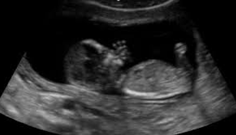
Once the baby is located and the fetal heartbeat is confirmed, the direction of the scan turns to allow the mother to witness the life growing inside of her. As imagers, we are on Holy Ground as we are literally watching the baby being knitted in its mother’s womb right before our eyes. Let us take time to revel in the wonder with her as we are able to show her the baby’s features, including the beating heart. This is the most important time of the scan as the mother is able to see her baby for the first time. The baby’s feet and hands, along with the profile, are impactful images for her and her support system to see. These are perhaps the required images in imaging life.
After the mother has seen her baby and the baby’s features have been shown, it is important to document the required information regarding the exam.
If apregnancy is visualized, the following images should be documented. A transvaginal exam may be performed if information is not obtainable transabdominally.
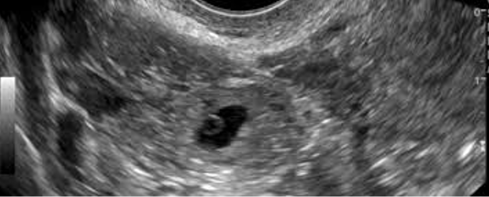
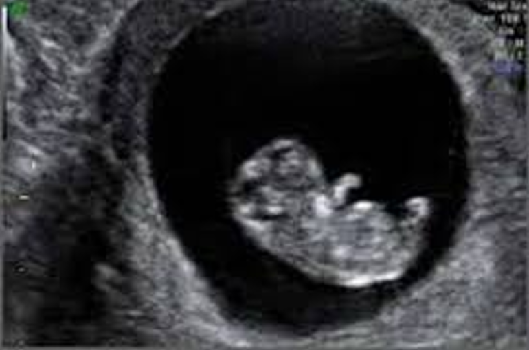
- A longitudinal or transverse image documenting the location of the gestational sac within the endometrial cavity within the uterus.
- An m-mode image documenting the baby’s heart rate.
- A gestational measurement. (Mean Sac Diameter, Crown-Rump Length or Femur Length)
- A copy of the worksheet or report from the machine with the EDD and Gestational Age.
If, while completing the systematic survey, an area of concern is visualized, documentation of that area should also be included as directed by each specific PHMC Policy and Procedure per the Medical Director.
If an ectopic pregnancy is visualized, the following images should be documented.
- A longitudinal or transverse image documenting an empty endometrium and (if present) a pseudosac.
- A longitudinal or transverse image documenting the location of the ectopic pregnancy.
- Yolk sac (if present)
- An m-mode image documenting the baby’s heart rate. (if present)
- A gestational measurement, if available, or image of an adnexal mass. (Mean Sac Diameter or Crown-Rump Length)
- A longitudinal or transverse image documenting any presence of free fluid.
- A copy of the worksheet or report from the machine with the Gestational Age.
Policies and procedures should indicate the images required for the Limited Obstetrical Ultrasound Exam. They may vary greatly from each PHMC's Medical Director based on their personal preference. For some PHMCs, the preference may be dependent on the type of permanent image created.
As an imager, it is to be noted that whether the image is documented as a permanent image or not does lessen the responsibility of the imager to duly image systematically for each scan. The beauty of ultrasound is its dynamic ability to image life by creating literal images as the transducer is being angled, leaving the responsibility of imaging thoroughly with the imager. Therefore, each image created is to be evaluated.
Imaging Life is such a gift, a privilege, and a responsibility. However, it is imperative that we remember the purpose behind the exams within pregnancy help. The purpose, the why, is to give her the information she needs at that moment in time to make the decision she needs to make. That information is extremely limited when compared to the complete and more in-depth ultrasound studies that the baby will receive during prenatal care if its mother chooses life.
Let us not get too caught up in the technicalities of trying to perform a complete study that hasn’t been ordered, while sacrificing precious time that could be spent imaging the baby for the mother to see.
It truly is so much more than just a scan.