by Tammy Stearns, MS, RDMS, RVT, RT(R), FSDMS, FAIUM, LAS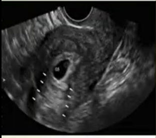
Often when one hears the term artifact, there is the reactionary thought of how to correct it, eliminate it and make it disappear. However, in ultrasound certain artifacts can be useful to the imager and contribute to the information gleaned from the exam. If utilized well, certain artifacts help to determine the composition of the structure, the technical features of the exam, and the direction in which the imager should follow while imaging.
Basic Properties of Sound
The physical properties of sound, how it traverses the body, and ultimately contribute to the composition of the image on the ultrasound screen, is daily utilized by imagers to help determine the structures being imaged. The basic parameters of imaging with sound include the transmission of waves by a transducer which react with the body’s tissues to create images based on acoustic impedance, reflection, scattering, and attenuation. The type of interaction that occurs is dependent on the stiffness and density of the structures that it encounters. As the sound beam travels through the body, it weakens in accordance with the composition of the structures in which it traverses. The portion of the beam that bounces back to the transducer is then converted into a level of white, black, or various shades of gray, dependent on the intensity of the received sound beam in comparison to the sound beam that was transmitted. Weaker signals are blacker while strong signals are brighter or white.
Helpful Artifacts
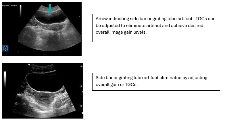
The Side Lobe or Grating artifact can often be found in the full bladder, which is necessary to scan a transabdominal pelvis. The interface of the bladder wall and the fluid within the bladder creates conditions that cause the sound beam to create what appears to be white or echogenic (white) lines across the anterior portion of the bladder. They are typically faint and can extend beyond the bladder wall. They are not consistent in their presence or appearance as the structure is imaged in two planes, nor do they remain as the bladder is emptied or when the patient changes positions, thus confirming they are artifact by nature. While they can be degrading to the overall appearance of the image, they provide a reference as to how to adjust the overall gain of the image. This can be accomplished by adjusting the overall gain or moving the TGCs inward. By knowing that they are simply artifacts, the imager can utilize them as a guide to darken the image by decreasing the gain until they are no longer visible.
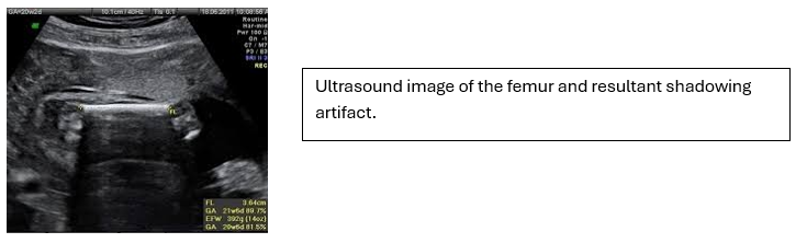
Shadowing occurs when an object has a significant attention coefficient that results in the sound beam hitting the object and instead of passing through the object, the sound beam bounces directly back. This results in the structure posterior to the object to not be imaged or displayed on the ultrasound screen due to lack of information gained. An example would be the femur length. The sound beam will travel to the femur, a hard bone with high levels of stiffness and density, hit the bone and bounce back to the transducer. Everything posterior to the femur is not imaged due to the sound beam not being able to pass by the femur itself. This provides the imager with the information that this structure has a significant level of stiffness and density confirming, most often, that it is either bone or a significant calcification. This is not an artifact that needs to be minimized or eliminated yet is utilized to help in the imaging process. If it is necessary to image the structures directly posterior to the structure that are obscured by the shadowing, scanning at a different angle or through a different sonographic window can be helpful.
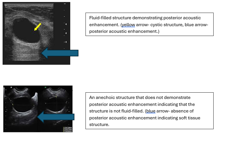
Posterior Acoustic Enhancement is an artifact that provides information regarding the cystic nature of the structure. As the sound beam travels through the body, it is attenuated at different levels dependent on the structure through which it is traveling. The attenuation rate is determined by the level of stiffness and density of the structure. Whereas shadowing is caused by the great level of attenuation of high level of stiffness and density in bone, a structure that is cystic allows the sound beam to travel through with less attenuation than the soft tissue that surrounds it which results in the area directly posterior to the structure to be brighter than the adjacent soft tissue. This allows the imager to differentiate between structures as being soft tissue masses or cystic (fluid-filled). Certain masses can appear to be cystic due to their hypoechoic or almost anechoic (black) appearance, however, without demonstrating posterior acoustic enhancement (brightness or white appearance directly posterior), it can be determined that they are not fluid-filled because of the attenuation that has traveled through the structure is the same as the adjacent soft tissue. However, a structure with the same appearance as a soft tissue mass can be determined to be cystic or have significant cystic components as it demonstrates posterior acoustic enhancement or brightness posterior to the structure.
By recognizing and utilizing the information obtained with artifacts that are helpful to the imager can determine whether structures are cystic, soft tissue, or bone. The artifacts can also help the imager determine the level of darkness necessary regarding the overall image appearance on the ultrasound screen. While there are artifacts that can lead to image degradation and need to be eliminated, the artifacts that offer key information are invaluable to the imaging process.