by Tammy Stearns, MS, RDMS, RVT, RT(R), FSDMS, FAIUM, LAS
One of the most important responsibilities of an imager performing a limited obstetrical ultrasound is determining whether the gestational sac is implanted in the appropriate location within the uterus. Although identifying a gestational sac may initially appear straightforward, accurately confirming its correct location within not just the uterus but within the endometrial tissue requires more than simply locating a small, fluid-filled structure on the ultrasound screen. It requires an understanding of maternal anatomy, proper transducer orientation, systematic scanning techniques, and the critical-thinking skills necessary to distinguish the normal findings of gestational sac location from those requiring further medical evaluation. For imagers performing limited obstetrical ultrasound in the Pregnancy Help Medical Clinic (PHMC), this distinction is particularly important. Documenting the presence of the gestational sac is not the same as confirming that the gestational sac is located in the correct location.
Understanding the Anatomy of the Uterus
Before identifying a gestational sac, the imager must identify the uterus. The uterus consists of three primary parts: the fundus, body, and cervix. The fundus is the rounded, superior portion of the uterus, located above the entrances of the fallopian tubes. The uterine body extends downward toward the lower uterine segment and cervix. In early pregnancy, normal implantation generally occurs within the decidualized endometrium of the upper two-thirds of the uterine cavity. Additionally, the uterus may be anteverted, retroverted, or positioned at various angles within the maternal pelvis. These anatomical variations influence how the uterus appears during transabdominal scanning and the location of the gestational sac in comparison to the maternal pelvis. Therefore, it is important to first identify the uterus and then identify the location of the gestational sac in comparison to the uterus.
A systematic transabdominal scan begins with the transducer positioned longitudinally at the level of the pubic symphysis angling toward the client’s head utilizing the urinary bladder as an acoustic window. The imager will utilize the hyperechoic linear endometrial stripe to lengthen out the uterus connecting the cervix, body and fundus in their entirety. Once the uterus is imaged in its entirety with the endometrial cavity imaged, the gestational sac can be identified.
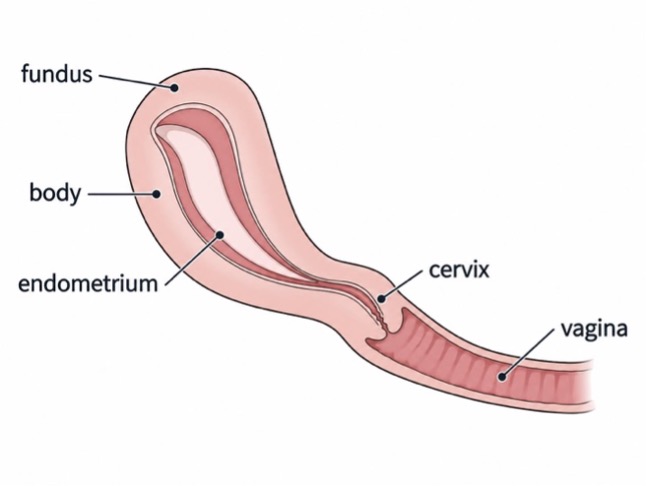 Diagram of the uterus longitudinal demonstrating the vagina, cervix, body and fundus.
A useful critical-thinking question at this point is:
“Can I demonstrate where this structure is located in relation to the entire uterus?”
If the answer is no, additional scanning is necessary before making conclusions about pregnancy location.
The endometrium provides one of the most important anatomical landmarks when evaluating early pregnancy. During pregnancy, the endometrium undergoes decidual changes, becoming thicker and more echogenic. A normally implanted gestational sac develops within this decidualized endometrium. On ultrasound, an early gestational sac typically appears as a small, round or oval fluid collection surrounded by an echogenic rim demonstrating a double decidual reaction. It is important to note that the presence of a fluid collection alone does not definitively establish an intrauterine pregnancy. The imager must evaluate whether the structure is located within the expected endometrial region and whether its relationship to the uterine cavity is consistent with normal implantation. Visualization of a yolk sac within a normally located intrauterine fluid collection provides definitive evidence of an intrauterine gestational sac.
After confirming the gestational sac, the imager should evaluate its position relative to the uterine fundus. The imager should determine whether the gestational sac is positioned within the upper uterine endometrial region rather than the lower uterine segment, cervix, or an abnormally lateral implantation site. It is important to note that a gestational sac may appear slightly eccentric within the endometrium while still representing a normally located pregnancy. However, a sac positioned far laterally near the uterine cornu requires careful evaluation because an interstitial ectopic pregnancy can resemble a fundal intrauterine pregnancy. The critical distinction is whether the gestational sac is completely surrounded by endometrium or separated from the endometrial cavity by myometrial tissue. The endometrial tissue will appear echogenic or white whereas the myometrial tissue will appear hypoechoic or darker than the endometrial.
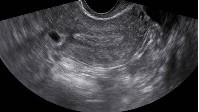 Ectopic Interstitial pregnancy located outside of the endometrial cavity.
After confirming the gestational sac longitudinally, the sonographer should rotate the transducer approximately 90 degrees to obtain a transverse view. The uterus should then be systematically evaluated from the cervix through the fundus. This helps establish the right-to-left relationship of the gestational sac within the uterine cavity. A structure appearing centrally located in the sagittal plane may appear unusually lateral in the transverse plane. Similarly, an apparently fundal structure may not actually be within the endometrial cavity. A single image rarely provides enough anatomical information to confidently establish implantation location. Both sagittal and transverse views should support the same interpretation.
It should be recognized that early pregnancies may present particular challenges during transabdominal ultrasound. Small gestational sacs, large maternal body habitus, uterine position, inadequately filled bladder, and equipment limitations can affect visualization. It should be noted that an excessively full bladder may distort uterine anatomy, while an inadequately filled bladder may limit the acoustic window. The imager must recognize when technical limitations prevent adequate assessment. Importantly, failure to visualize a normally located gestational sac does not establish that an ectopic pregnancy is present. Likewise, the presence of an intrauterine fluid collection does not automatically exclude an ectopic pregnancy. When the pregnancy location remains uncertain, the client requires timely evaluation through the established medical referral process, which may include diagnostic transvaginal ultrasound and serial quantitative hCG measurements depending on possible dating of the pregnancy.
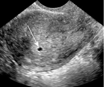 Ultrasound image demonstrating the correct location of an early gestational sac demonstrating a double decidual reaction.
Before concluding that a gestational sac is normally located, the imager should mentally review several questions:
- Have I identified the entire uterus and its anatomical orientation?
- Can I clearly identify the fundus and endometrial cavity?
- Is the suspected gestational sac located within the expected upper uterine endometrial region?
- Have I confirmed its relationship to the endometrium in both sagittal and transverse planes?
- Is a yolk sac or embryo visible to support identification of a definite pregnancy?
- Are there unusual findings or technical limitations requiring further evaluation?
These questions encourage deliberate observation rather than assumption.
Any patient experiencing significant pelvic pain, vaginal bleeding, dizziness, syncope, or other concerning symptoms requires appropriate medical assessment. Severe pain, shoulder pain, fainting, or signs of instability warrant emergency evaluation because ectopic pregnancy complications can be life-threatening.
Confirming the gestational sac location during early pregnancy requires much more than the simple finding of a round, dark circle on an ultrasound screen. It requires the imager to understand uterine anatomy, recognize the endometrial cavity, identify the fundus, and systematically evaluate the suspected gestational sac in multiple imaging planes. Most importantly, it requires knowing when the available images do not provide sufficient information. The goal of critical thinking in limited obstetrical ultrasound is not simply to find the pregnancy. It is to understand what has been visualized, recognize what remains uncertain, and respond appropriately to protect the patient.
This helps answer the questions that the expectant mother has regarding her pregnancy to help her make the decisions that she needs to make.