by Tammy Stearns, MS, RDMS, RVT, RT(R), FSDMS, FAIUM, LAS
Prior to scanning a Limited Obstetrical Exam, part of the preparation of the exam is preparing for what will be imaged. Most often, a normal intrauterine pregnancy will be imaged with the heart rate measured with m-mode, gestational age noted by gestational sac measurement or crown rump length, and all the essential images allowing mom to experience the reality of life growing inside of her. At times, however, other scenarios are encountered to include normal variants and abnormal conditions that shed a different light on the pregnancy. Some of these scenarios can be of minimal significance, while some can have life-altering consequences. The presence of some of the scenarios can even be both dependent on the variables of the factors that present during the scan. The presence of free fluid is one of these scenarios. Minimal free fluid in the pelvis can be explained by physiological changes, whereas complex or significant free fluid in the pelvis can be the first indication that conditions needing further investigation are present.
Prior to the ultrasound exam, while obtaining the client’s history, it is important to note any history of pelvic fullness or discomfort in the pelvic region. Some may describe the sensation as needing to have a bowel movement. Depending on the amount of free fluid present, left shoulder pain may be present due to irritation of the diaphragm. This is especially evident during deep inhalation. Symptoms such as these give indication of the presence of free fluid prior to the actual scan.
Clear fluid presents as anechoic, or black, within the pelvis when imaged by ultrasound. Fluid can be classified as simple (completely black) or complex (having echoes, septations or debris within). Fluid can also be described as minimal or significant depending on the amount present. It is not enclosed within a capsule, so it fills the empty spaces within the most dependent locations of the pelvis pending the position of the client. Since most clients are scanned lying on their back, the most dependent location in the pelvis is the Pouch of Douglas (rectouterine cul-de-sac) located posterior to the uterus. Morrison’s Pouch, located between the right kidney and the liver, is also considered a common dependent imaging location and can be imaged to verify the presence of free fluid.
 |
|
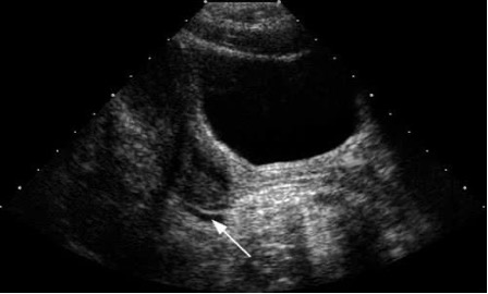
Minimal amount of free fluid as indicted by the arrow. |
The cause of free fluid varies. In the non-pregnant female, the presence of a small amount of free fluid can be a normal physiological finding related to ovulation, menstruation, or normal peritoneal fluid. Larger amounts can indicate pathological issues like ruptured ovarian cysts, pelvic infections, or inflammation. Small amounts of fluid may be present at the time of ovulation when the dominant cyst releases the egg. The dominant cyst then becomes the corpus luteum and secretes progesterone which causes thickening of the endometrium in anticipation of a pregnancy. The corpus luteum will typically regress by the next cycle if a pregnancy does not occur. Pelvic Inflammatory Disease (PID) can also cause large amounts of complex fluid within the pelvic region due to inflammation. Debris and septations within the fluid may be present.
 |
|
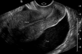
Significant free fluid with echoes posterior to the uterus. |
Free fluid present with a positive pregnancy test can be indicative of physiological changes due to pregnancy or can be indicative of pathological conditions. Dependingdependent on the amount and sonographic appearance of the presenting fluid, this can indicate the cause of the free fluid. Normal physiologic causes include small, trace amounts of fluid that naturally pool in the pelvic cavity typically posterior to the uterus or due to the corpus luteum cyst that ruptured or is leaking small amounts of fluid. After the fertilized egg has been implanted into the thickened endometrium, hCG will be released, indicating the presence of a pregnancy. This signals the corpus luteum to continue secreting progesterone to support the early pregnancy until the placenta takes over, at about ten to twelve weeks gestation, maintaining the thickened endometrium and the cessation of menstruation. Most cysts are harmless, painless, and disappear on their own by the second trimester. They can, however, rupture or leak minimal amounts of fluid causing the presence of free fluid. A minimal amount of free fluid may also be present at the time of ovulation when the dominant corpus luteum cyst releases the egg.
 |
|
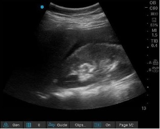
Morrison’s Pouch, located between the right kidney and liver, demonstrating the presence of free fluid. |
The most common pathologic causes of free fluid encountered during pregnancy can be due to an ectopic pregnancy or pelvic infection. Free fluid seen during an ectopic pregnancy is almost always blood (hemoperitoneum) leaking from the implantation site. As the fertilized egg grows outside the uterus, most commonly in the fallopian tube, it stretches and eventually tears local blood vessels or ruptures the organ entirely, causing blood to pool in the pelvic cavity. The appearance of the fluid is important to note. Small, simple fluid, defined as a tiny amount of clear fluid in the pouch of Douglas can sometimes be a normal finding from recent ovulation. However, with a positive test it should be noted, especially if an intrauterine pregnancy has not been confirmed. Complex or echogenic fluid that contains echoes, debris, or blood clots strongly indicates the presence of an ectopic pregnancy requiring urgent medical care. Just as with the non-pregnant exam, Pelvic Inflammatory Disease can cause large amounts of complex fluid within the pelvic region. Debris and septations within the fluid maybe present.
 |
|
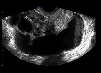
Free fluid with debris present with PID. |
The presence of free fluid on the ultrasound exam is not a definitive that pathology is present, however, it does require that the amount and appearance of the free fluid be evaluated to decide if next steps need to be taken. A minimal amount of fluid that is simple and clear may simply be physiologic changes. A significant amount of fluid requires additional imaging and investigation. The appearance of the fluid from simple to complex further indicates that the fluid may be of pathological origin. The type of documentation of the free fluid will be dependent on the direction of each Pregnancy Help Medical Clinic’s Medical Director and the approved medical policies and procedures.